by Robert W. Chandler, MD, MBA, Daily Clout:

Dr. Arne Burkhardt is one of eight international pathologists, physicians and scientists who were asked to perform a second autopsy, requested by friends and family of the deceased who were not satisfied with the results of the first autopsy.
Thirty autopsies and three biopsies were evaluated; 15 cases with routine histopathology (Step 1), three with advanced methods (Step 2), and some of the remaining 15 are included as illustrative cases.
The Step 1 group included eight women and seven men aged 28-95 (average 69).
TRUTH LIVES on at https://sgtreport.tv/
Death occurred seven days to 180 days following the first or the second Spike-Mediated Gene Therapy (SMGT) with COMIRNATY in eight, Moderna in two, AstraZeneca in two, Janssen in one and Unknown in two.
Place of death was known in 17 cases:
- Nine Non-hospital: five at home, one on the street, one in a car, one at work, one in an elder care facility
- Eight Hospital: four ICU, four died having been in hospital less than two days
Special stains were used to identify Spike and Nucleocapsid Proteins, with the following differential:
- COVID-19 (C-19) = + Spike + Nucleocapsid.
- SMGT = + Spike – Nucleocapsid.
Causation by SMGT: Very probable in five cases, probable in seven, unclear in two and no connection in one.
Lesions were on multiple organs including: Brain, Heart, Kidney, Liver, Lungs, Lymph Node, Salivary Gland, Skin, Spleen, Testis, Thyroid and Vascular.
Lymphocyte Infiltration, present in 14 of 20 cases (70%), was a common feature and involved multiple organs. Case 19 had at least five different organs involved. CD3+ Lymphocytes were dominant.
The Vascular System was targeted by Lymphocyte Infiltration in seven (35%) of the cases and included sloughing endothelium, destruction of the vessel wall, hemorrhage and thrombosis.
A condition called Lymphocyte Amok was described by Dr. Burkhardt: Lymphocyte accumulation in non-lymphatic organs and tissues that might develop into lymphoma.
Five cases of unknown foreign material in blood vessels were identified. The favored explanation for origin of this material was aggregated Lipid Nanoparticles (LNPs).
Multiple pathologic processes were involved: Apoptosis, Coagulopathy, Clotting/Infarction, Infiltration/Mass Formation, Inflammation, Lysis, Necrosis and Neoplasia.
Röltgen, et al. https://www.cell.com/cell/fulltext/S0092-8674(22)00076-9 found that COVID-19 depleted Lymphatic Germinal Centers (LGCs) whereas SMGT stimulated them, suggesting a possible origin of “Hunter/Killer” CD3+ Lymphocytes that are attracted to certain tissues, particularly the vascular system.
An expanded program of autopsy following SMGT is recommended in order to further understand the actions of SMGTs and to help formulate new treatments for the constellation of pathology associated with such drugs.
Burkhardt Group Conclusions:
- Histopathologic analyses show clear evidence of vaccine-induced autoimmune-like pathology in multiple organs.
- That myriad adverse events deriving from such auto-attack processes must be expected to very frequently occur in all individuals, particularly following booster injections.
- Beyond any doubt, injection of gene-based COVID-19 vaccines place lives under threat of illness and death.
- We note that both mRNA and vector-based vaccines are represented among these cases, as are all four major manufacturers.
Histopathology
This report is the first in a series in which harms from the Lipid Nanoparticle (LNP) Messenger Ribonucleic Acid (mRNA) therapeutics and other Spike-mediated products will be examined from the point of view of the pathologist, a medical doctor that studies specimens obtained from removal of tissue from living persons, bulk resection or biopsy, or after death. Such examinations make or confirm a diagnosis and provide a basis to determine causation of tissue mass or cause of death. Histopathology refers to the study of abnormal tissues.
Tissues are examined using careful inspection of specimens with the naked eye followed by examination by light microscopy employing a variety of different stains to highlight important features of cells, tissues and organs. A common stain used is hematoxylin and eosin, H & E for short, which stains nuclei blue, cytoplasm pink or red, collagen fibers pink and muscles red.
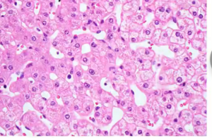
Many of the photomicrographs in this and subsequent articles will have been stained with H & E. Pathologists display sections prepared with H & E along with the magnification used, such as 40 times (40X) or 100 times (100X) magnification.
Immunohistochemical Stains for COVID-19 and Spike-Mediated Therapy
Special stains are vital to the identification of certain histopathology, such as cases involving Spike-mediated therapeutics. These examinations require special stains as outlined by Dr. Arne Burkhardt whose specimens and lecture notes will be the subject of this first report in a series.
Dr. Burkhardt discusses below the immunohistochemistry staining techniques necessary to differentially diagnose cell/tissue damage/organ from COVID-19, SARS-CoV-2 or something else, as well as specific cell types of interest such a T-lymphocytes and monocytes:
Immunohistochemistry to detect vaccine-induced spike protein expression.
Prof. Dr. A. Burkhardt
- Use anti-SARS-COV-2 spike protein/S1 antibodies to test for presence of spike protein in tissue samples. Always include myocardium and spleen tissue samples.
- If spike protein is detected, use anti-nucleocapsid antibody to examine expression of SARS-COV-2 nucleocapsid: presence of nucleocapsid indicates viral “breakthrough” infection, absence of nucleocapsid supports vaccine-induced spike protein expression.
- Perform positive and negative controls using vaccine-transfected and non-transfected cell cultures.
Differential Staining to identify CD3 and CD68 cells and to Differentiate COVID-19 from Spike-Inducing Drugs
COVID-19 = Spike stain + Nucleocapsid stain +
LNP/mRNA = Spike stain + Nucleocapsid stain -:
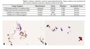
- Spike, red arrow. b. Nucleocapsid, red arrow.
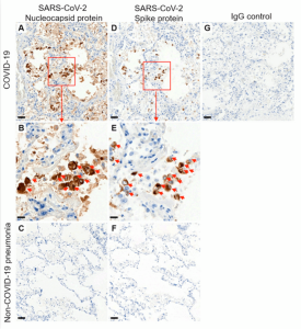
Without these special stains and without an exhaustive search of the specimens, no autopsy should be considered complete.
The internet is an excellent source for examples of both normal and pathological cells, tissues and organs.
A useful guide to have available when looking at the photomicrographs to follow is the Histology Guide at: https://histologyguide.com/.
This internet tool can be used to examine normal histology and compare it to the histopathology seen in Dr. Burkhardt’s slide deck which has been integrated with the transcription from his lecture on February 5, 2022.
Autopsy-Histology-Study on Vaccination-Associated Complications and Deaths
(Dr. Burkhardt’s slide deck was reproduced here and was integrated with the text derived from notes compiled from the voice recognition transcript with limited editing for readability with no intention to make substantive changes.)
Understanding Vaccine Causation Conference – February 5, 2022
World Council for Health
Dr. Arne Burkhardt
Pathologist
Reutlingen, Germany
“Dr. Arne Burkhardt, born in 1944 in Germany, a pathologist with more than 40 years diagnostic and teaching experience at the Universities of Hamburg, Bern, and Tübingen. He is the author of more than 150 original publications in international journals, currently engaged in autopsy studies of persons dying after taking the Covid vaccine.” Shabhan Palesa Mohamed

https://worldcouncilforhealth.org/multimedia/uvc-arne-burkhardt/
Dr. Burkhardt: I think it’s very, very, important to have an international communication on this subject, because last year in May, April, I was confronted with some relatives of persons who died after vaccination. And I tried to establish a national registry of these persons dying.
And I tried to get autopsies done in these persons, but the national associations of pathology here didn’t reply to this request by myself. So, when relatives continued to ask me, “Where can I get some solution to this problem?” finally, I said, “I can examine these organ probes that have been taken during autopsy, and we can try to get some other pathologists sent.”


